
Arthroscopic Removal Of Loose Bodies In The Knee
Minimally invasive removal of loose fragments to reduce knee pain and improve joint movement
Loose bodies are small, loose fragments of cartilage or bone that float around the knee joint. They can occur for various reasons in osteoarthritis, and rheumatoid arthritis. Other causes include fractures, trauma, bone and cartilage inflammation, and benign tumours of the synovial membrane.
A loose body in the knee can cause a sensation of locking, making it difficult to move or fully extend the joint. They can cause pain, swelling, and locking, and catching of the joint and may be found in individuals who participate in sportsince they are more susceptible to fractures and other sports injuries.
DIAGNOSIS OF LOOSE BODIES IN THE KNEE

To diagnose a loose body, Dr Martin will conduct a thorough evaluation, including a review of your medical history and a physical examination.
The following diagnostic tests may also be performed:
X-ray (Radiograph):This is usually the first test performed when diagnosing a loose body. An X-ray uses radiation to produce a black-and-white image of the joint, highlighting any bone or cartilage fragments. Since most loose bodies contain some bone or cartilage, they are typically visible on X-rays.
Computed Tomography (CT) Scan:A CT scan combines X-ray technology with computer processing to create detailed cross-sectional images of the joint. This test provides a higher-definition image than a standard X-ray, allowing for better visualization of the loose bodies and their exact location.
Magnetic Resonance Imaging (MRI):Unlike X-rays and CT scans, an MRI uses electromagnetic radio waves instead of radiation. During the MRI, you will be placed inside a scanner that generates detailed images of the joint. This test is particularly useful for detecting loose bodies that do not contain bone and would not be visible on other imaging tests.
Arthrography: In some cases, arthrography may be used to obtain a more detailed view of the joint. This involves injecting a contrast dye into the affected area and taking an X-ray. The dye highlights the soft tissues and cartilage, providing a clearer image. Although CT scans and MRIs are often preferred, arthrography can still be useful for imaging smaller joints, such as the wrist.
SURGERY FOR LOOSE BODIES REMOVAL IN THE KNEE
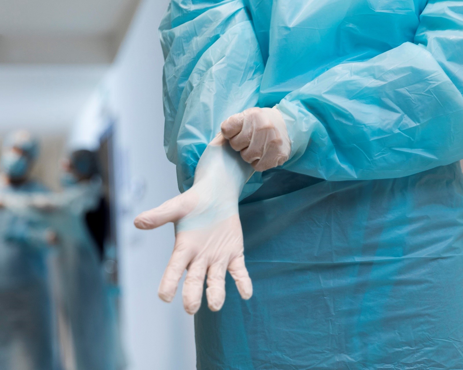
Dr Martin typically performs loose body removal in the knee under either local or general anaesthesia, depending on individual specifics and patient needs.
ARTHROSCOPIC LOOSE BODY REMOVAL
The most common method for removing loose bodies in the knee is through arthroscopic surgery.
This minimally invasive procedure involves the following steps:
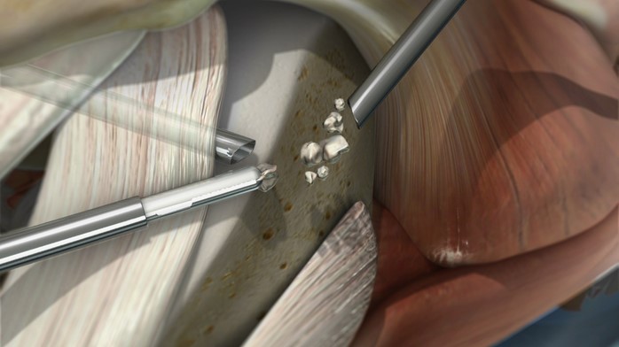
Incisions and Arthroscope Insertion: Dr Martin makes two to three small keyhole incisions around the knee. These incisions allow access to the knee joint and the insertion of an arthroscope—a small camera that provides a clear view of the knee’s interior.
Saline Infusion: Through one of the portals, Dr Martin fills the knee with a sterile saline solution. This helps expand the knee joint, providing better visibility and space to manoeuvre surgical instruments.
Locating and Removing Loose Bodies: Using specialised tools, such as a grabber, Dr Martin locates and removes the loose bodies from the knee joint. The arthroscope allows precise navigation and extraction of these fragments.
Closing the Incisions: After all loose bodies are removed, the small incisions are closed with sutures or surgical staples, completing the procedure.
ARTHROTOMY FOR LOOSE BODY REMOVAL
In cases where the loose bodies are too large to be removed arthroscopically, Dr Martin may perform an arthrotomy.
This procedure involves:
Larger Incisions: Making larger incisions to provide better access to the knee joint.
Direct Removal: Directly removing the loose bodies that cannot fit through the smaller portals used in arthroscopy.
ADDITIONAL LOOSE BODY REMOVAL TECHNIQUES
Suction and Graspers: Depending on the size and location of the loose bodies, Dr Martin may use a suction tip or a small needle to grasp and remove the fragments.
Mechanical Burr or Resector: For loose bodies located in hard-to-reach areas or those that need to be broken down, a mechanical burr or resector may be used to break them into smaller, manageable pieces, which the body can then degrade naturally.
Fixation with Screws or Pins: In cases where loose bodies are caused by fractures or osteocartilaginous lesions, they may be fixed in place using screws or pins.
Partial Synovectomy: If the loose body is caused by a benign tumour of the synovial membrane, Dr Martin may perform a partial synovectomy, which involves removing part of the knee joint lining.
POST-SURGICAL CARE – LOOSE BODIES REMOVAL FROM THE KNEE
Undergoing surgery for the removal of loose bodies from the knee is a significant step towards regaining joint function and relieving discomfort. Post-surgical care is crucial to ensure a successful recovery and restore mobility. Dr Martin provides comprehensive post-operative care guidelines to support your healing process and help you return to your normal activities as safely and quickly as possible.
Immediate Post-Surgery Care
Pain Management: Pain relief is a critical component of your recovery. You will be prescribed pain medication to manage any post-operative discomfort. Follow the prescribed dosage and schedule to maintain effective pain control.
Applying ice packs to the knee can help reduce swelling and alleviate pain. Use ice packs for 15-20 minutes every 1-2 hours for the first few days post-surgery.
Wound Care:Keep the surgical site clean and dry. Your knee will be covered with a sterile dressing immediately after surgery. Change the dressing as instructed by Dr Martin or his team. Watch for signs of infection, such as increased redness, swelling, warmth, or discharge from the incision site, and report any concerns immediately.
Elevation and Rest:Elevate your knee above heart level as much as possible to reduce swelling and promote healing. Use pillows to support your leg while resting or sleeping. Avoid putting weight on the operated knee initially. Use crutches or a walker as directed to assist with mobility and prevent strain on the knee.
Early Rehabilitation (First Few Weeks)
Physiotherapy: Begin physiotherapy as soon as recommended by Dr Martin. Early mobilisation and guided exercises are essential to regain range of motion, strengthen the muscles around the knee, and prevent stiffness. Your physiotherapist will provide a tailored exercise program, focusing on gentle stretching, bending, and straightening exercises to improve flexibility and strength.
Activity Restrictions: Avoid high-impact activities, heavy lifting, and any movements that may stress the knee. Gradually increase your activity level based on your comfort and the guidance of your healthcare team.
Follow-Up Appointments: Attend all scheduled follow-up appointments with Dr Martin. These visits are crucial for monitoring your healing progress, adjusting your treatment plan if necessary, and addressing any concerns.
Intermediate Rehabilitation (Weeks 3-6)
Continued Physiotherapy: Your physiotherapy program will evolve to include more challenging exercises aimed at building strength, improving balance, and enhancing joint stability.
Activities may include stationary cycling, leg presses, and resistance exercises tailored to your recovery stage.
Gradual Weight-Bearing: Begin to gradually increase weight-bearing on the operated knee as tolerated. Your physiotherapist will guide you on how to transition from crutches to walking without assistance.
Swelling Management:Continue to use ice packs as needed to manage swelling, especially after physiotherapy sessions. Compression bandages or knee sleeves can also help control swelling and provide support.
Advanced Rehabilitation (Weeks 6 and beyond)
Strength and Conditioning: Engage in more advanced strengthening exercises, such as squats, lunges, and step-ups, to rebuild muscle strength and endurance.
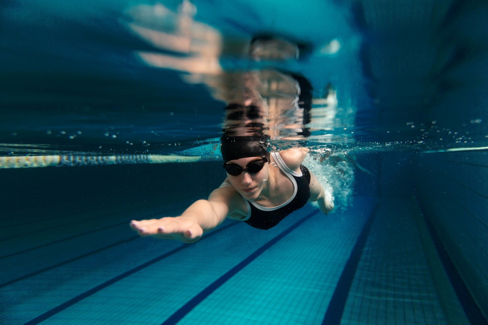
Cardiovascular activities like swimming or using an elliptical machine can enhance overall fitness without placing undue stress on the knee.
Sport-Specific Training:If you are an athlete or involved in physical activities, your rehabilitation program will include sport-specific drills to prepare you for a safe return to your chosen activities. Drills may involve agility training, plyometrics, and controlled jumping and landing exercises to ensure your knee can handle the demands of your sport.
Psychological Readiness:Mental recovery is as important as physical healing. Work with your physiotherapist and Dr Martin to build confidence in your knee’s strength and stability. Address any fears or anxieties about returning to physical activities and take gradual steps to regain your pre-injury level of performance.
Long-Term Care and Maintenance
Ongoing Exercise: Maintain a regular exercise routine to keep the muscles around your knee strong and flexible. This helps prevent future injuries and supports overall knee health.
Incorporate low-impact activities such as walking, cycling, or swimming into your daily routine.
Regular Check-Ups:Schedule periodic follow-up appointments with Dr Martin to monitor your long-term recovery and address any ongoing concerns. Stay proactive about your knee health and seek medical advice if you experience any new or recurring symptoms.
Lifestyle Adjustments:Consider lifestyle modifications to reduce the risk of future knee injuries. This may include maintaining a healthy weight, using proper techniques during physical activities, and wearing appropriate footwear.
By following these detailed post-surgical care guidelines and working closely with Dr Sam Martin and your healthcare team, you can achieve a successful recovery from loose body removal surgery and return to your normal activities with improved knee function and comfort.